Protein Surface Mimetics
CHEMISTRY, BIOCHEMISTRY, PHARMACOLOGY, PROTEIN SURFACE MIMETICS, GPCR REGULATION, ENZYME REGULATION, ANTI-INFLAMMATORY RESEARCH, ANTI-INFECTIVES RESEARCH, ANTI-CANCER RESEARCH, NEURODEGENERATIVE RESEARCH, CARDIOVASCULAR RESEARCH
Our group has been developing small molecules that can structurally and functionally mimic small discrete elements of protein secondary structure (e.g. strands, helices, loops, multiple motifs), as these are often crucial components in protein-protein interactions and are responsible for mediating biological activities. Examples of target protein surfaces include:
a) Beta strands are commonly recognized in substrates and inhibitors of enzymes called proteases (e.g. Tyndall et al, Chem. Rev. 2005) and kinases, and in beta sheet formation in proteins such as those resulting in neurodegenerative diseases (Harrison et. al 2007).
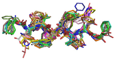
Figure 1. Multiple inhibitors of HIV-1 protease showing the common extended beta strand backbone conformation.
b) Turn motifs in peptides and proteins that activate G Protein-Coupled Receptors (e.g. Tyndall et al, Chem. Rev. 2005).
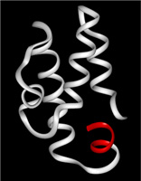
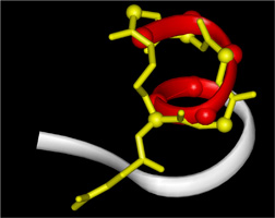
Figure 2. Helical turn motif (red) at the C-terminus of C5a (white) is mimicked by the cyclic peptide "3D53" or "PMX53" (yellow) designed in our lab.
c) Alpha helices which we can readily stabilise within short peptides in water using constraints such as lactam bridges (e.g. Shepherd et al, J. Am. Chem. Soc. 2005, Shepherd et al, Angewandte Chemie 2004) or or metal clips (e.g. Kelso et al, J. Am. Chem. Soc. 2000; Kelso et al, J. Am. Chem. Soc. 2004) These are the smallest known alpha helices and the first examples of single alpha helical turns ever reported.
Figure 3: Molecular Dynamics simulations of alpha-helix mimetics - double click on the two structures to make them move Left: An unconstrained alpha-helix rapidly becomes unstructured in dynamics simulations and in water, whilst a sidechain-constrained bis-lactam bridge analogue remains helical over a 2 nanosecond MD run and in water.
d) Discontinuous loops of proteins which we have mimicked by grafting peptide loops onto macrocyclic scaffolds (e.g. Singh et al, J. Am. Chem. Soc. 2005)
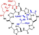
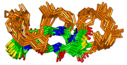
Figure 4: Two and Three Loop Mimetics Loops stabilized by parallel or antiparallel positioning on a macrocyclic scaffold - note how the scaffold directs loops from the same face, enabling their coordinated interactions with receptors. double click on the central structure to see it rotate (e.g. Singh et al, Org. Lett. 2002 Singh et al, Org. Lett. 2006/)